Patella Tendon Repair Rehab Protocol: A Comprehensive Guide
This comprehensive guide details a structured rehabilitation pathway following patella tendon repair surgery, spanning from initial post-operative care through return to activity.
It outlines progressive phases, emphasizing pain management, range of motion, strengthening, and functional restoration—typically over 3-6 months.
The protocol incorporates insights from recent research (2017-2024) regarding biomechanics, osteopathic correction, and optimal post-surgical recovery timelines.
Understanding Patella Tendon Repair
Patella tendon repair addresses tears or ruptures of the tendon connecting the kneecap (patella) to the shinbone (tibia). These injuries commonly occur in athletes participating in jumping sports, but can also result from falls or direct trauma. The patella tendon is crucial for extending the knee, enabling activities like walking, running, and jumping.
Surgical intervention becomes necessary when conservative treatments fail to alleviate pain and restore function. Repair techniques vary, often involving suture anchors to reattach the torn tendon to the bone. Understanding the biomechanics of the patellofemoral joint – the interaction between the patella and femur – is vital for successful rehabilitation, as highlighted in recent biomechanical analyses (Yurova, 2024).
Post-surgical rehabilitation is a critical component of recovery, aiming to restore strength, range of motion, and stability. A well-defined protocol, like the one detailed here, guides patients through progressive phases, minimizing complications and optimizing functional outcomes. Early rehabilitation, even within the ICU or shortly after surgery (Belkin, 2022), is increasingly recognized for its benefits.
Indications for Patella Tendon Repair
Patella tendon repair is typically indicated when a tear or rupture significantly impairs knee function and doesn’t respond to conservative management. Complete tendon ruptures, often presenting with inability to extend the knee, necessitate surgical intervention. Partial tears causing persistent pain and weakness, especially in active individuals, are also strong candidates for repair.
Specific indications include pain localized to the inferior pole of the patella, swelling, bruising, and difficulty with weight-bearing activities. Athletes involved in jumping or pivoting sports frequently require repair to restore their pre-injury level of function. Dysplastic changes within the patellofemoral joint, potentially requiring medial patellofemoral ligament plasty alongside tendon repair (Suchilin, 2021), can also influence the decision.
Diagnosis usually involves a physical examination, assessing range of motion, stability, and pain provocation tests. Imaging studies, such as MRI, confirm the extent of the tear and rule out other associated injuries. The goal of surgical repair is to restore the tendon’s anatomical attachment, allowing for normalized biomechanics and a return to desired activity levels.
Surgical Techniques in Patella Tendon Repair
Surgical approaches for patella tendon repair vary depending on the tear’s location and severity. Common techniques involve direct repair, utilizing sutures to re-attach the torn tendon to the patella. In cases of significant tissue damage, tendon augmentation with allograft or autograft tissue may be necessary to reinforce the repair.
Arthroscopic techniques are increasingly utilized, offering minimally invasive access and visualization. Open repair may be preferred for complex tears or when addressing concurrent patellofemoral joint instability, potentially requiring medial patellofemoral ligament (MPFL) reconstruction (IA Suchilin, 2021). Bone-tendon-bone autografts are frequently employed for ACL reconstruction, influencing rehabilitation protocols.
The surgeon’s preference and the specific characteristics of the injury dictate the chosen method. Post-operatively, the repaired tendon is typically protected with a cast or brace to allow for initial healing. Careful attention to surgical technique and subsequent rehabilitation are crucial for optimal outcomes and a successful return to function.
Phase 1: Immediate Post-Operative Phase (0-2 Weeks)
Initial focus centers on pain and edema control, utilizing immobilization and cryotherapy. Weight-bearing is restricted, and gentle range of motion exercises begin cautiously.
Early quadriceps activation is initiated to prevent atrophy.
Weight-Bearing Restrictions

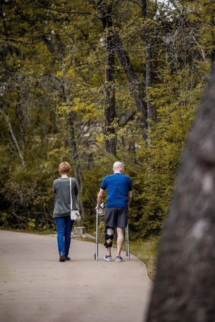
Following patella tendon repair, strict adherence to weight-bearing restrictions is crucial for optimal healing. Initially, touch-down weight-bearing is permitted, meaning the operated leg can lightly touch the ground for balance during ambulation, but no weight is loaded through it. This phase typically lasts for the first 0-2 weeks post-surgery.
Progression to partial weight-bearing, utilizing crutches, occurs as tolerated and guided by the surgeon’s protocol and pain levels. The goal is to gradually increase weight-bearing capacity, aiming for 25-50% body weight by the end of week two. Regular assessment of the surgical site and pain response is essential to prevent overloading the repair.
Full weight-bearing is generally not permitted until at least 6-8 weeks post-operatively, contingent upon adequate quadriceps strength, pain control, and radiographic evidence of tendon healing. Precise criteria for advancing weight-bearing status will be determined by the surgical team, considering individual patient factors and the specific repair technique employed. Consistent monitoring and adherence to these guidelines are paramount to avoid complications and ensure a successful recovery.
Pain and Edema Management
Effective pain and edema control are fundamental during the initial post-operative phase (0-2 weeks). Pharmacological interventions, including prescribed analgesics, are utilized to manage pain levels, allowing for participation in rehabilitation exercises. Ice application, for 15-20 minutes several times daily, is crucial for reducing swelling and inflammation.
Elevation of the leg above heart level, whenever possible, further aids in edema reduction. Compression bandages are applied to minimize swelling and provide support. Gentle range of motion exercises, initiated early, help to prevent stiffness and promote lymphatic drainage, contributing to edema control.
Patient education regarding pain management strategies, including activity modification and proper icing techniques, is essential. Monitoring pain levels using a visual analog scale (VAS) guides progression of rehabilitation. Addressing pain proactively ensures optimal patient compliance and facilitates a smoother recovery process. Early intervention is key to preventing chronic pain and complications.
Range of Motion Exercises (ROM) ― Phase 1
Initiating gentle range of motion (ROM) exercises is paramount in Phase 1 (0-2 weeks) to prevent stiffness and promote healing. Heel slides are introduced early, progressing as tolerated, aiming for pain-free movement. Ankle pumps are performed frequently to enhance circulation and reduce swelling, indirectly aiding knee mobility.
Patellar mobilization, performed by a therapist, gently restores patellar movement, preventing adhesions. Emphasis is placed on achieving full knee extension as a primary goal, while flexion is progressed cautiously, respecting pain limits. Active-assisted ROM, utilizing a towel or strap, assists with flexion and extension.
Exercises are performed within a pain-free range, avoiding any stress on the repaired tendon. Regular monitoring of ROM is crucial to track progress and adjust the exercise program accordingly. Patient education emphasizes the importance of consistent, gentle movements to optimize recovery and prevent complications. Avoidance of forceful stretching is critical during this initial phase.
Early Quadriceps Activation
Early quadriceps activation is crucial, despite weight-bearing restrictions, to minimize muscle atrophy and facilitate functional recovery. Isometric quadriceps sets are initiated immediately post-operatively, focusing on tightening the thigh muscles without moving the knee joint. These are held for 5-10 seconds, repeated frequently throughout the day.
Neuromuscular electrical stimulation (NMES) may be employed to augment quadriceps activation, particularly if voluntary contraction is difficult. Straight leg raises (SLR) are introduced cautiously, ensuring minimal stress on the repair, and performed with the knee fully extended. Emphasis is placed on maintaining proper form and avoiding compensatory movements.
Co-contraction of the hamstrings is encouraged to provide stability during SLR exercises. Progressive increases in hold time and repetitions are implemented as tolerated. Patient education stresses the importance of gentle, controlled contractions to avoid disrupting the healing tendon. Monitoring for pain and swelling is essential to guide exercise progression.

Phase 2: Intermediate Post-Operative Phase (2-6 Weeks)
This phase focuses on continued pain and edema control, progressive knee flexion, and initiating strengthening exercises, typically 2-6 weeks post-surgery, guided by rehabilitation goals.
Continued Minimization of Pain and Edema
Managing post-operative pain and edema remains a priority during this intermediate phase (2-6 weeks). Consistent application of ice packs for 15-20 minutes several times daily is crucial. Elevation of the leg above heart level, whenever possible, further aids in reducing swelling.
Pharmacological interventions, as prescribed by the physician, should be adhered to diligently. Gentle range of motion exercises, initiated in Phase 1, are continued to prevent stiffness and promote fluid drainage. Kneecap taping, a physiotherapy technique, can provide support and reduce discomfort.
Monitoring for signs of increased pain, redness, or warmth is essential, as these may indicate infection. Patient education regarding activity modification and proper body mechanics is vital to prevent re-injury and optimize healing. The goal is to achieve a pain level that allows for comfortable participation in rehabilitation exercises, facilitating progressive loading of the patellar tendon.
Progressive Knee Flexion Goals
Achieving progressive knee flexion is a cornerstone of rehabilitation between weeks 2 and 6 post-surgery. Initial goals focus on attaining 90 degrees of flexion by week 3, and 120 degrees by week 4. These targets are not rigid and will be individualized based on patient tolerance and pain levels.
Active-assisted range of motion (AAROM) exercises, utilizing a towel or continuous passive motion (CPM) machine, are employed to gently increase flexion. Heel slides and seated knee bends are also beneficial. Avoidance of forceful or passive stretching is crucial to protect the healing tendon.
Regular monitoring of flexion angles is essential to track progress and adjust the rehabilitation program accordingly. Pain should be carefully monitored during flexion exercises; any significant increase warrants modification of the activity; The ultimate aim is to restore full, pain-free range of motion, preparing the knee for more advanced strengthening exercises.
Strengthening Exercises ― Phase 2

Phase 2 strengthening (weeks 2-6) prioritizes restoring quadriceps strength while protecting the repaired patella tendon. Isometric quadriceps sets are initiated early, progressing to short-arc quads and straight leg raises. These exercises focus on activating the muscle without placing excessive stress on the tendon.
Gentle hamstring curls and hip abduction/adduction exercises are incorporated to address surrounding muscle weakness. Emphasis is placed on controlled movements and avoiding any activities that cause pain or swelling. Resistance bands are gradually introduced to increase the challenge.
Closed kinetic chain exercises, such as mini-squats (within a pain-free range), begin cautiously. The goal is to improve strength and proprioception. Monitoring for any signs of tendon irritation is crucial, and exercises should be modified or discontinued if necessary. Progressive overload is key, but must be carefully managed.
Patellar Mobilization Techniques
Patellar mobilization is crucial during Phase 2 (weeks 2-6) to prevent stiffness and restore normal patellar tracking. Gentle, graded mobilizations are performed by a qualified therapist to address any restrictions in patellar movement—superior/inferior, medial/lateral.
These techniques aim to improve the gliding mechanics of the patella within the femoral groove, preventing adhesions and promoting optimal biomechanics. Soft tissue mobilization around the patella and quadriceps muscle belly is also implemented to release tension and improve flexibility.
The therapist will assess the patella’s mobility and tailor the techniques accordingly, ensuring patient comfort and avoiding any exacerbation of pain. Mobilizations are typically combined with range of motion exercises to maximize their effectiveness. Regular assessment and adjustments are vital throughout this phase, contributing to a successful recovery.
Phase 3: Advanced Strengthening Phase (6-12 Weeks)
This phase focuses on building strength and endurance with closed and open kinetic chain exercises, alongside proprioceptive training and cardiovascular conditioning for optimal function.
Closed Kinetic Chain Exercises
Closed kinetic chain (CKC) exercises are paramount during the advanced strengthening phase (6-12 weeks) post-patella tendon repair, as they promote functional movement patterns and enhance stability. These exercises involve the foot being fixed on a surface, allowing for simultaneous co-contraction of muscles around the knee and hip.
Examples include progressively loaded squats (starting with partial squats and advancing to full squats), leg presses, lunges (forward, reverse, and lateral), and step-ups. Resistance can be increased through the use of weights, resistance bands, or incline adjustments. The focus should be on maintaining proper form and avoiding compensatory movements.
CKC exercises are beneficial because they minimize stress on the repaired tendon while maximizing muscle activation. They also improve proprioception and neuromuscular control, crucial for regaining functional activities. Progression is guided by pain levels and objective measures of strength and stability. Careful monitoring ensures appropriate loading and prevents re-injury. The goal is to build strength and endurance in a manner that closely mimics real-life movements.
Open Kinetic Chain Exercises
Open kinetic chain (OKC) exercises, initiated during the advanced strengthening phase (6-12 weeks), complement closed kinetic chain exercises in the patella tendon repair rehabilitation protocol. OKC movements involve the foot being free in space, isolating the quadriceps muscle group.
Common examples include leg extensions, hamstring curls, and seated calf raises. Initially, these exercises are performed with minimal resistance, focusing on controlled movements through a full range of motion. Resistance is gradually increased as strength improves, utilizing weight stacks or resistance bands.
OKC exercises are valuable for targeting specific muscle groups and addressing any remaining strength deficits. However, they must be implemented cautiously, as they can place greater stress on the patellofemoral joint. Proper form and controlled speed are essential to prevent pain or re-injury; The integration of OKC and CKC exercises provides a comprehensive approach to restoring full knee function and preparing the patient for a return to activity.
Proprioceptive Training
Proprioceptive training is a crucial component of the advanced strengthening phase (6-12 weeks) in patella tendon repair rehabilitation, focusing on restoring the knee’s ability to sense its position in space. Following surgery, proprioception is often impaired, increasing the risk of re-injury.
Exercises include single-leg stance with eyes open and closed, wobble board activities, and balance beam walking. These activities challenge the patient’s balance and coordination, stimulating the neuromuscular system to regain control. Progression involves incorporating dynamic movements, such as reaching or tossing a ball while maintaining balance.
The goal is to improve joint stability and reduce the likelihood of giving way. Integrating proprioceptive exercises with functional activities helps translate improved balance into real-world movements. Consistent practice is essential for long-term gains, enhancing the patient’s confidence and ability to return to desired activities safely and effectively.
Cardiovascular Conditioning
Cardiovascular conditioning is integrated into the rehabilitation protocol, beginning gradually in Phase 3 (6-12 weeks) and continuing through the return-to-activity phase. Maintaining overall fitness is vital, even with weight-bearing restrictions and limitations in lower extremity function post-surgery.
Initial activities focus on non-impact modalities like stationary cycling with minimal resistance, upper body ergometry, and swimming (avoiding breaststroke kick). As strength and range of motion improve, low-impact activities such as elliptical training and walking can be introduced, carefully monitoring for pain or swelling.
The objective is to improve endurance, reduce cardiovascular deconditioning, and support overall recovery. Gradually increasing the duration and intensity of cardiovascular exercise is key. This component complements strengthening and proprioceptive training, contributing to a holistic rehabilitation approach and preparing the patient for the demands of functional activities and eventual return to sport.

Phase 4: Return to Activity Phase (3-6 Months)
This final phase prioritizes functional exercises and sport-specific training, ensuring a safe return to desired activities. Criteria-based progression is essential, guided by objective testing and minimizing re-injury risk.
Functional Exercises
Functional exercises bridge the gap between controlled rehabilitation and the demands of daily life or sport. These movements emphasize integrated, multi-planar activities, mimicking real-world scenarios. Initially, focus on bilateral exercises like step-ups, squats (progressing depth cautiously), and lunges, ensuring proper form and pain-free execution.
Progression involves unilateral variations, increasing the challenge to the repaired patella tendon. Agility drills, such as cone drills and shuttle runs, are introduced to enhance dynamic stability and proprioception. Plyometric exercises – jumping and hopping – are incorporated cautiously, starting with low-impact options and gradually increasing intensity.
Key considerations include maintaining proper biomechanics throughout each exercise, avoiding compensatory movements, and monitoring for any signs of pain or swelling. The goal is to restore functional movement patterns, enabling a smooth and confident return to activity. Regular assessment and adjustments to the exercise program are crucial, based on individual progress and tolerance. This phase often requires 12-16 weeks of consistent effort.
Sport-Specific Training
Sport-specific training represents the final phase before returning to full activity, meticulously replicating the movements and demands of the individual’s chosen sport. This phase builds upon the foundation of strength, power, and agility established in previous stages. For example, a runner will progress through interval training, gradually increasing speed and distance, while a jumper will focus on plyometrics tailored to jumping mechanics.
Exercises should mimic game-like situations, incorporating cutting, pivoting, and rapid changes in direction. The intensity and volume are carefully monitored, avoiding overexertion and minimizing the risk of re-injury. Neuromuscular control is a key focus, ensuring the athlete can react quickly and efficiently to unexpected movements.
This stage requires close collaboration between the physical therapist and the athlete’s coach to design a program that is both challenging and safe. Gradual progression is paramount, with regular assessments to track progress and adjust the program as needed. Successful completion of this phase prepares the athlete for a confident and sustainable return to sport.
Criteria for Return to Sport
Returning to sport following patella tendon repair necessitates meeting specific, objective criteria to minimize re-injury risk. These benchmarks extend beyond simply achieving pain-free movement and encompass functional performance indicators. Firstly, full, symmetrical range of motion is essential, alongside no reported pain during or after activity.
Strength testing must demonstrate at least 90% limb symmetry index (LSI) for key muscle groups – quadriceps, hamstrings, and calf muscles. Functional hop tests, including single-leg hop for distance, triple hop, and crossover hop, should also achieve >90% LSI. Agility tests, like the T-test and shuttle run, are crucial for assessing dynamic stability and control.
Furthermore, the athlete must demonstrate satisfactory performance in sport-specific drills without compensatory movement patterns. Psychological readiness is also vital; the athlete must feel confident and prepared to return. A thorough assessment by the rehabilitation team, considering all these factors, is paramount before granting clearance for full participation.

Long-Term Considerations
Sustained success relies on adherence to a continued maintenance program, addressing potential complications like re-tear or stiffness. Proactive measures and consistent exercise are key.
Long-term monitoring and preventative strategies are vital for optimal function and minimizing future issues post-surgery.
Potential Complications
Several complications can arise following patella tendon repair, necessitating vigilant monitoring during rehabilitation. Re-tear of the repaired tendon, though infrequent, represents a significant concern, potentially requiring revision surgery. Infection, while minimized with prophylactic antibiotics, remains a risk, presenting with increased pain, redness, and swelling.
Stiffness and limited range of motion are common, often addressed through aggressive early mobilization and progressive ROM exercises. Patellar maltracking, or improper kneecap movement, can occur, potentially leading to pain and cartilage damage. Nerve injury, affecting sensation around the knee, is a rare but possible complication.
Chronic pain syndromes can develop, requiring multidisciplinary management. Quadriceps weakness may persist, impacting functional recovery. Adverse reactions to hardware, if used in the repair, are also possible. Careful adherence to the rehabilitation protocol and prompt reporting of any concerning symptoms are crucial for mitigating these risks and optimizing long-term outcomes.
Preventative Measures
Proactive strategies are essential to minimize complications and optimize recovery post-patella tendon repair. Strict adherence to weight-bearing restrictions and the prescribed rehabilitation protocol is paramount, particularly during the initial phases. Meticulous wound care, including regular cleaning and monitoring for signs of infection, is crucial.
Early and consistent pain and edema management, utilizing ice, elevation, and analgesics, helps facilitate participation in rehabilitation. Progressive strengthening exercises, guided by a physical therapist, rebuild quadriceps and hamstring strength, supporting patellar stability. Proprioceptive training enhances neuromuscular control, reducing re-injury risk.
Maintaining optimal nutrition supports tissue healing. Avoiding high-impact activities until cleared by the surgical team prevents undue stress on the repair. Regular communication with the healthcare provider regarding any concerns or setbacks allows for timely intervention and protocol adjustments, ensuring a safe and effective return to function.
Importance of Continued Maintenance
Long-term success following patella tendon repair hinges on a commitment to ongoing maintenance. Even after achieving full return to activity, a consistent exercise program is vital to preserve strength, flexibility, and proprioception. Regular quadriceps and hamstring strengthening prevents muscle imbalances that could compromise patellar tracking.
Continued proprioceptive exercises refine neuromuscular control, enhancing joint stability and reducing the risk of re-injury. Incorporating low-impact cardiovascular conditioning maintains overall fitness without excessive stress on the repaired tendon. Paying attention to biomechanics during activities, and addressing any movement patterns that place undue strain on the knee, is crucial.
Periodic check-ins with a physical therapist can identify and address any subtle deficits before they escalate. Adopting a proactive approach to injury prevention, including proper warm-up and cool-down routines, safeguards the repair and ensures long-term functional capacity.


0 Comments